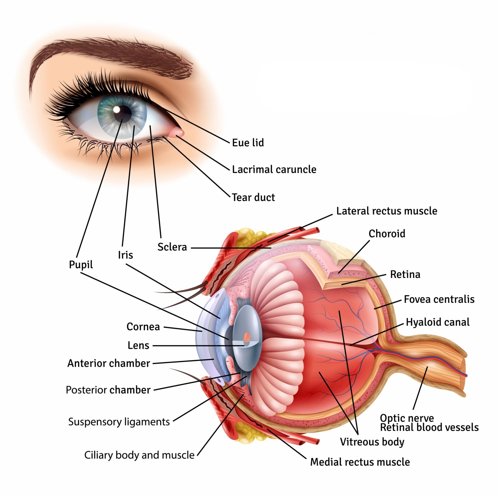
The anatomy of the human eye is a marvel of biological precision. This extraordinary organ contains structures that work in perfect harmony to transform light into the vivid world we see. From the transparent cornea that first focuses incoming light to the retina's millions of photoreceptors that convert it to electrical signals, each component serves a crucial role in the visual process. Discover how this intricate system allows you to perceive depth, distinguish millions of colors, and adapt instantly to changing environments—all in milliseconds.
The Remarkable Engineering of Human Vision
Understanding the anatomy of the human eye reveals a masterpiece of biological engineering. Your eyes are extraordinary biological cameras, capturing millions of visual signals every second before transmitting them to your brain for interpretation. This intricate system allows you to perceive depth, distinguish approximately 10 million colors, and adapt instantly to changing light conditions.
According to research published in Medical News & Life Sciences, humans possess binocular vision—our two eyes work together to create a single combined image with remarkable depth perception. This visual processing system is considered one of the most complex and efficient biological systems in nature.
Let’s explore the fascinating structures that make vision possible and understand how they impact your everyday sight.
External Eye Structures: Your First Line of Defense
Protective Elements
Orbit The eye rests within a protective bony socket called the orbit. Six specialized extraocular muscles attach to the eye within this cavity, enabling precise movements in multiple directions—side-to-side, up-and-down, and rotational movements that allow you to track moving objects smoothly.
Eyelids These protective covers perform several critical functions:
Blocking light during sleep
Distributing tears across the eye surface
Blinking reflexively to protect against foreign objects
Maintaining optimal moisture levels
Eyelashes and Eyebrows These seemingly decorative features serve crucial protective purposes:
Filtering dust and airborne particles
Redirecting sweat away from the eyes
Reducing glare from overhead light sources
Providing sensory warning when objects approach too closely
Tear Production System
Tear Glands Located primarily in the upper eyelid, these specialized glands produce tears that:
Maintain consistent eye lubrication
Contain antibacterial enzymes that fight infections
Wash away irritants and foreign particles
Provide essential nutrients to the cornea
The Visible Eye: What We See in the Mirror
The Window to Vision
Sclera The white, visible portion of your eye is the sclera—a tough, fibrous tissue covering approximately 83% of the eye’s surface. This durable outer layer:
Maintains the eye’s shape
Protects internal structures
Provides attachment points for the extraocular muscles
Resists internal and external pressure
Conjunctiva This thin, transparent membrane covers the sclera and lines the inside of your eyelids. The conjunctiva:
Contains small blood vessels that may enlarge when irritated (causing redness)
Produces mucus that helps lubricate the eye
Forms a protective barrier against pathogens
Helps prevent foreign objects from reaching sensitive eye tissues
Cornea The clear, dome-shaped surface at the front of your eye is the cornea. This remarkable structure:
Provides approximately 70% of the eye’s focusing power
Contains no blood vessels, maintaining transparency
Has one of the highest densities of pain receptors in the body
Self-heals minor abrasions remarkably quickly
Iris The colored portion of your eye is genetically determined and as unique as your fingerprint. The iris:
Controls light entry by adjusting pupil size
Contains melanin—the same pigment that determines skin color
Consists of two muscle groups that work in opposition to precisely control pupil diameter
Pupil The black circular opening at the center of the iris is the pupil. This adjustable aperture:
Expands to 4-8mm in low light conditions
Contracts to 2-4mm in bright environments
Can change size almost instantaneously
Appears black because light entering is mostly absorbed by internal eye tissues
Internal Eye Structures: The Image Processors
Light Management System
Lens Located behind the iris, this flexible, crystalline structure:
Fine-tunes focus after light passes through the cornea
Changes shape through accommodation—becoming thicker for near vision and thinner for distance vision
Gradually hardens with age, contributing to presbyopia (difficulty focusing on close objects)
Filters some harmful UV radiation
Ciliary Muscles These tiny circular muscles surrounding the lens:
Contract to thicken the lens for near vision
Relax to flatten the lens for distance vision
Work continuously throughout your waking hours
Often fatigue when focusing on close work for extended periods
Vitreous Humor This transparent, jelly-like substance fills the space between the lens and retina:
Maintains the eye’s spherical shape
Transmits light to the retina with minimal distortion
Contains fine fibers that help suspend the lens in place
Consists of 99% water with collagen fibers and hyaluronic acid
Image Reception Center
Retina This light-sensitive layer at the back of the eye:
Contains approximately 120 million rod cells (for night and peripheral vision)
Houses roughly 6 million cone cells (for color vision and detail)
Transforms light signals into electrical impulses
Is actually an extension of brain tissue
Macula Located near the center of the retina, this specialized region:
Occupies only about 5% of the retina but processes most of your visual information
Contains the highest concentration of cone cells
Provides the sharp central vision needed for reading, driving, and facial recognition
Has a slight yellow pigmentation that filters harmful blue light
Optic Nerve This bundle of approximately 1 million nerve fibers:
Transmits visual information from the retina to the brain
Creates a natural blind spot where it connects to the retina
Carries visual signals at speeds approaching 250 mph
Connects to multiple brain regions for comprehensive visual processing
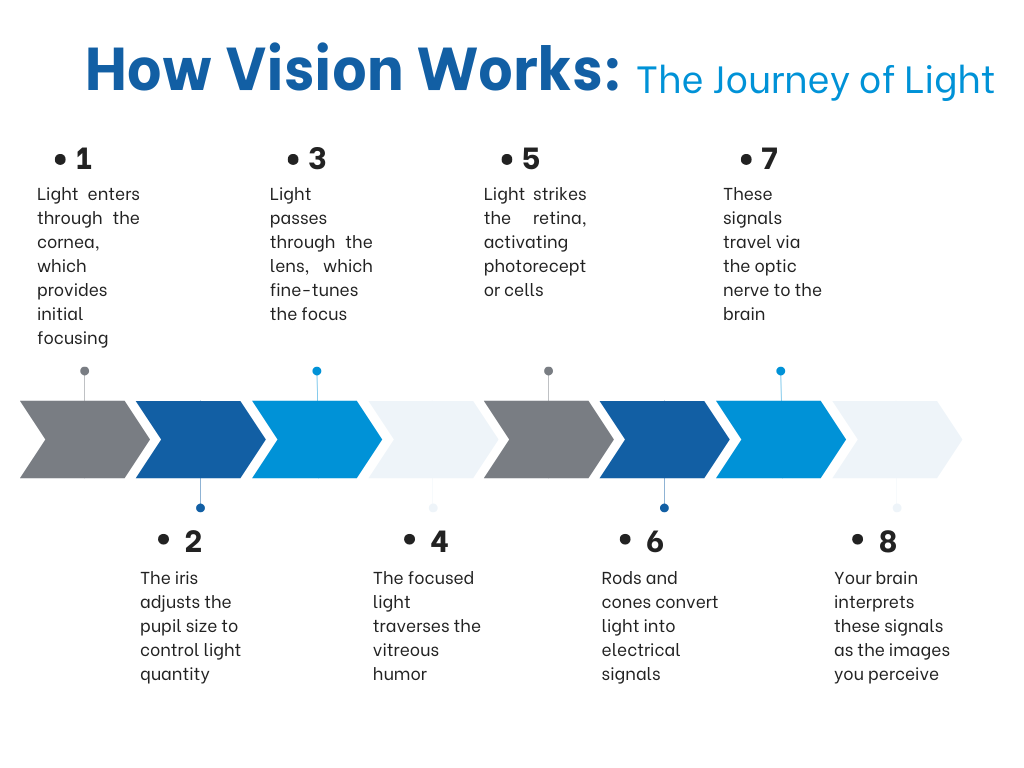
How Vision Works: The Journey of Light
When you look at an object, light travels through a precise sequence:
Light enters through the cornea, which provides initial focusing
The iris adjusts the pupil size to control light quantity
Light passes through the lens, which fine-tunes the focus
The focused light traverses the vitreous humor
Light strikes the retina, activating photoreceptor cells
Rods and cones convert light into electrical signals
These signals travel via the optic nerve to the brain
Your brain interprets these signals as the images you perceive
This entire process occurs in milliseconds, allowing you to perceive motion smoothly and react to visual changes instantly.
Common Vision Challenges and Solutions
When any part of this intricate system experiences changes, vision can be affected. Common conditions include:
Presbyopia: Age-related difficulty focusing on close objects
Vision Correction Options Traditional solutions include eyeglasses and contact lenses, which adjust how light enters the eye and focuses on the retina. However, many people are turning to surgical options like LASIK for more permanent vision correction.
LASIK: A Modern Solution for Vision Correction
LASIK (Laser-Assisted In Situ Keratomileusis) eye surgery offers several advantages:
Quick procedure: Typically takes less than 30 minutes for both eyes
Minimal recovery time: Most patients return to normal activities within 24-48 hours
High success rate: Over 96% of patients achieve their desired vision
Reduced dependency: Minimizes or eliminates the need for glasses or contacts
Long-lasting results: Benefits typically last for many years or even decades
Is LASIK Right for You?
When considering LASIK, consultation with an experienced eye care professional is essential. They can assess your specific needs and determine if you’re a suitable candidate based on: